
3D-Printed Lung Models Enhance Planning for Pulmonary Malformation Procedures
Written with artificial intelligence.
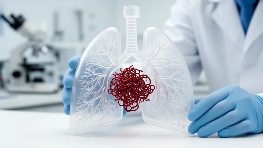
A study published in 3D Printing in Medicine demonstrated that patient-specific 3D-printed models of pulmonary arteriovenous malformations (PAVMs) were successfully created for six patients, aiding interventional radiologists in procedural planning. The models not only improved anatomical understanding but also enhanced patient education regarding their conditions.
Study Overview
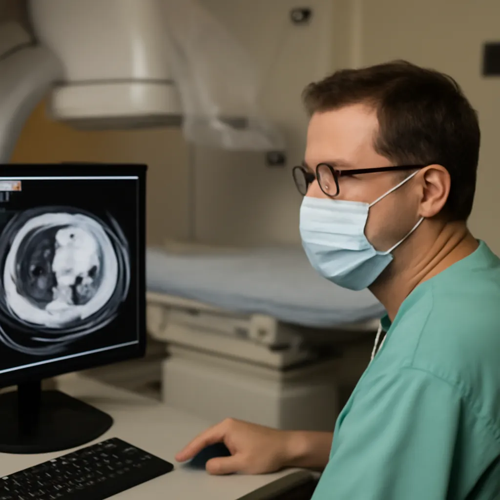
Interventional radiologists often rely on two-dimensional CT scans to navigate complex vascular structures like pulmonary arteriovenous malformations (PAVMs). A recent feasibility study, conducted by researchers from the University of Pittsburgh Medical Center, the University of California San Francisco, and Washington University School of Medicine, explored the use of 3D-printed models for better procedural planning. The study involved six patients with eight PAVMs, with all models successfully fabricated using advanced printing techniques.
The Challenge of PAVMs
PAVMs involve abnormal connections between pulmonary arteries and veins, bypassing the capillary network. This condition can lead to serious complications, including strokes and low oxygen levels. The standard treatment is embolization, which is complicated by the anatomy of the malformations. Traditional imaging can make it difficult for interventionalists to accurately visualize and plan procedures, especially for complex cases.
3D Printing Methodology
The study utilized thin-slice CT angiography to create detailed digital models of each patient's vascular anatomy. These models were then fabricated using multimaterial printing, allowing for distinct visualization of arterial and venous structures. This approach ensures that the models can be consistently produced across different institutions, making it a viable tool for routine clinical use.
Clinical Implications
The use of these models led to a clearer understanding of the vascular anatomy before procedures, contributing to a 100% technical success rate in embolization attempts. Additionally, the models served as valuable educational tools for patients, converting complex medical concepts into tangible objects that facilitate informed consent and understanding. However, the study's authors caution that further research is needed to quantify the impact of these models on procedural efficiency and patient outcomes.
Conclusion
As 3D printing technology continues to advance, its applications in clinical settings are expanding. This study highlights the potential for patient-specific models to improve the planning and execution of difficult procedures in interventional radiology, underscoring the need for ongoing research to validate these findings and explore their broader implications in medical practice.