
3D Printed Lung Phantom Developed for Pulmonary Embolism Imaging
Written with artificial intelligence.
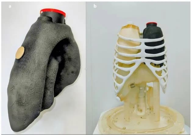
Researchers at the University Hospital of Brest in France have created a 3D printed lung model that realistically simulates a pulmonary embolism. This innovative phantom aims to enhance the accuracy of CT and nuclear medicine imaging by providing a reliable reference for measuring blood flow obstruction.
Introduction to the Lung Phantom
Researchers from the University Hospital of Brest in France have successfully 3D printed a fillable left lung model designed to simulate pulmonary embolism (PE). Utilizing a Formlabs Fuse 1+ selective laser sintering (SLS) system, this model serves as a reference for CT and nuclear medicine scans, allowing for enhanced measurement accuracy in diagnosing PE.
Construction and Design Details
The team began by scanning the healthy lung insert of a Kyoto Kagaku thorax phantom and combined this with anonymized lung segmentation data using MIM Software’s image processing tools. They specifically designed the embolism zone in the superior segment of the left lower lobe for optimal simulation. The model, measuring 14.3 × 24.1 × 10.4 cm, was printed in Nylon 12, taking 17 hours to complete. Notably, it was printed off-site due to the hospital's lack of a large-format SLS printer.
Performance Evaluation
In evaluations, radiologists and nuclear medicine physicians rated the images produced by the phantom as highly realistic across all imaging modalities tested. The phantom's total volume was recorded at 5.04% over the reference lung's volume, with the embolism compartment accounting for 7.6% of the internal volume. The model effectively simulates lung tissue density using polystyrene beads, which corresponded closely to real lung densities.
Cost and Future Applications
The estimated production cost for the phantom, assuming standard market rates, is €770, excluding the cost of the printer and post-processing unit. The model's design offers potential for future studies, particularly in comparing imaging methods for perfusion defects and enhancing quality control across different scans. Furthermore, the researchers note that this workflow could be adapted for other organs, making it a versatile tool for medical imaging research.
Conclusion
This research addresses a significant gap in physical references for pulmonary embolism imaging, moving beyond generic models to a task-specific phantom. With its ability to simulate PE accurately, the 3D printed lung phantom is set to improve diagnostic practices in nuclear medicine and CT imaging.